
Quantitative Retinal Imaging
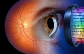
Current state-of-the-art imaging technologies can only detect structural damage from retinal disease when it is too advanced to be reversed. TNO’s patented Quantitative Retinal Imaging (QRI) device can uniquely detect oxygen distribution in even the smallest microvessels. This enables earlier and more quantitative diagnosis and disease monitoring, thereby improving treatment efficacy and patient outcomes.
Addressing the challenge
Reduced blood flow and oxygen availability in the retinal microvessels are the root cause of multiple retinal diseases, including diabetic retinopathy, glaucoma, retinal vein occlusion, and macular degeneration. Reduced flow leads to local retinal hypoxia. This can either damage retinal tissue directly or lead to the formation of fragile, leaky neovascularisation, which further damages the retina. State-of-the-art imaging systems, such as fundus cameras, laser scanning ophthalmoscopes, and OCT make images of the retina through the pupil. These only detect retinal diseases when irreversible structural damage has already occurred. Current retinal oximetry techniques can only detect oxygen levels in larger blood vessels, not in the microvessels.
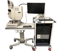
TNO’s patented Quantitative Retinal Imaging (QRI) technology is a multispectral, modulated retinal imaging device that uniquely facilitates the quantitative measurement of the light scattering and absorption properties in the retina. From this, TNO can extract novel biomarkers, including diagnostically relevant retinal microvascular oxygen saturation (StO2). Oxygen levels in even the smallest microvessels can be determined. This facilitates earlier, quantitative diagnosis and disease progression monitoring, allowing clinicians to make more informed treatment decisions and optimise patient care.
Expertise that differentiates
By combining their expertise in optical system design and multispectral light propagation in human tissue, TNO was able to develop and patent this novel approach to retinal disease detection. The QRI solution addresses the need for earlier and more quantitative detection of these irreversible retinal diseases, enabling faster treatment, improved progression monitoring, and better outcomes.
The use of modulated imaging to map oxygenation levels has already been demonstrated on skin tissue. The QRI system is the first known system to successfully demonstrate multicolour Spatial Frequency Domain Imaging (mcSFDI) within the retina. The novel biomarkers that were discovered can support research in ophthalmic, cardiovascular, and neurodegenerative diseases. As a market entry, mcSFDI could replace costly, invasive, and time-consuming fluorescence angiography procedures.
Applications that matter
For ophthalmologists in diagnostic centres, clinics, and university medical centres, earlier and more precise diagnosis means better patient outcomes. For patients, it means the vital preservation of healthy tissue and earlier recovery for those diagnosed with retinal diseases. For researchers, these novel biomarkers can have a significant impact on clinical and translational research into retinal, neurodegenerative, or cardiovascular disease.
After completing the experimental proof of concept, the QRI team has developed a research setup for the technology. The team has received ethical approval, and successfully created the first high-quality images from a healthy volunteer (TRL 3). In the next phase, TNO will conduct an exploratory clinical pilot (TRL 4) on a select number of people with retinal and neurodegenerative diseases. Based on the results of these trials, further development will take place.
Could QRI technology benefit your market position? Contact us for investment opportunities.
Contact us
-

Hidde Zittersteijn
Job title:Business Developer Biotech BoosterSpecialisation unknownHidde joined the TNO Ventures team as a business developer as part of the Biotech Booster “Nationaal Groeifonds” project. Driven by the potential of (bio)technology to improve societal challenges, Hidde aims to use his background to help these innovations achieve their impact in society. Hidde previously worked as a PhD candidate at the Leiden University Medical Center and as Business developer at the Leiden Bio Science Park foundation.
-
E-mail:E-mail Hidde
-
LinkedIn:Hidde on LinkedIn
-